
Part C preparation!
Onychomycosis , (etiology: dermatphytoses?)
Tinea corporis
Sprorotrichosis Pathophysiology: Infection with the dimorphic soil fungus S schenckii usually is acquired through cutaneous inoculation. The initial reddish, necrotic, nodular papule of cutaneous sporotrichosis generally appears 1-10 weeks after a penetrating skin injury. The lesion is a suppurating granuloma that consists of histiocytes and giant cells, with neutrophils that accumulate in the center and that are surrounded by lymphocytes and plasma cells.
The fungus spreads from the initial lesion along lymphatic channels, forming the chain of indolent nodular and ulcerating lesions that typifies the lymphocutaneous form of the disease.


Dermatophytoses

Tinea nigra
PSORIASIS

 Pathology
Skin biopsy shows acanthosis and parakeratosis, reflecting the increase in skin turnover. The granular layer is often absent. Polymorphonuclear abscesses may be seen in the upper epidermis. The epidermal rete ridges appear elongated and clubbed as they fold down into the dermis. Dermal changes include capillary dilatation surrounded by a mixed neutrophilic and lymphohistiocytic perivascular infiltrate.
Seborrheic keratosis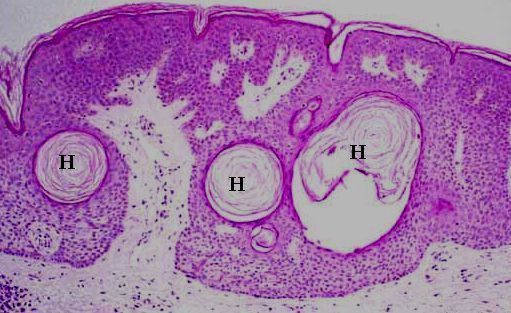
Halo nevus
squamous cell carcinoma of skin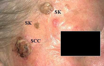
lichen planus
Melanoma 


Ovarian teratoma, mature 


Endometriosis

Cerebral Malaria
gametocyte of P.falciparum ?

Cutaneous larva migrans

Brain cysticercosis

posted by fooji @ 7:32 PM


 OS of the maxilla bone


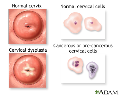
 CIN III
 Cervical invasive carcinoma


 Paget's disease of the breast
 ductal carcinoma in situ


 LCIS


 Invasive carcinoma
